Home
/ Anterior Neck Anatomy Diagram - Anterior Triangle Of The Neck Subdivisions Teachmeanatomy - .there are many head and neck anatomy rather than a detailed account for neurosurgery.
Anterior Neck Anatomy Diagram - Anterior Triangle Of The Neck Subdivisions Teachmeanatomy - .there are many head and neck anatomy rather than a detailed account for neurosurgery.
Anterior Neck Anatomy Diagram - Anterior Triangle Of The Neck Subdivisions Teachmeanatomy - .there are many head and neck anatomy rather than a detailed account for neurosurgery.. The anterior triangle is formed by the inferior border of the mandible, the anterior border of sternocleidomastoid and the sagittal plane in the midline of the. Head and neck anatomy human head face, muscles, people, head png. The two primary neck regions are the anterior cervical and posterior cervical triangles, which are found deep to the skin and subcutaneous tissue and contain several muscles, vasculature, and nerves. Neck and shoulder muscles diagram neck shoulder muscle anatomy shoulder muscle anatomy diagram anatomy. 3d interactive tutorials on the anatomy of the neck, including the anatomical organisation, musculature, larynx, pharynx, blood supply and innervation.
Vascular surgery tratamento disease vein, veins pituitary gland endocrine gland endocrine system anterior pituitary, brain, text, people png. Furthermore, the anterior triangle muscles are grouped depending on their position to the hyoid bone; Heads up assessing and activating cervical spine core muscles. On the left the normal contents of the carotid space and the derived pathology. 4 scalenus anterior muscles the scalenus anterior is an impt muscle of the lower part of the neck because of its relations with the impt structures in that region origin from.
Anterior Triangle Of The Neck from www.wesnorman.com The two primary neck regions are the anterior cervical and posterior cervical triangles, which are found deep to the skin and subcutaneous tissue and contain several muscles, vasculature, and nerves. Above to hyoid bone via thyrohyoid membrane, below to cricoid cart. Neck, in land vertebrates, the portion of the body joining the head to the shoulders and chest. In radiology, the 'head and neck' refers to all the anatomical structures in this region excluding the central nervous system, that is, the brain and spinal cord and their associated vascular structures and. Traditionally the anatomy of the infrahyoid neck has been subdivided into a group of surgical triangles whose borders are readily palpable bones it transverses the suprahyoid and infrahyoid neck into the anterior mediastinum. They pass between the posterior border of the sternocleidomastoid muscle and the upper border of the clavicle to drain into the external jugular veins in the posterior triangle of. Furthermore, the anterior triangle muscles are grouped depending on their position to the hyoid bone; Muscles of anterior neck and throat swallowing diagram.
Magnetic resonance imaging of the head and neck.
We hope you will use this picture in the study and. This article describes the anatomy of the head and neck of the human body, including the brain, bones, muscles, blood vessels, nerves, glands, nose, mouth, teeth, tongue, and throat. Muscles of anterior neck and throat swallowing diagram. The larynx is an important organ in the anterior neck. Muscles anatomy anterior neck muscle labeling anterior neck musculature neck muscle exercises neck skull muscle anatomy neck area anatomy neck muscles side view neck muscle pain head and neck muscle anatomy model neck muscles blank face muscle anatomy worksheet deep. The anterior and posterior triangles. On the left the normal contents of the carotid space and the derived pathology. Below this a thorough knowledge of anatomy and anatomical variations of the head and neck is essential to avoid or assess complications arising from tracheotomies. Lateral neck triangle the boundaries are the posterior border of the sternocleidomastoid the anterior border o. 3 write short notes on: Instant anatomy is a specialised web site for you to learn all about human anatomy of the body with diagrams, podcasts and revision questions. 3d interactive tutorials on the anatomy of the neck, including the anatomical organisation, musculature, larynx, pharynx, blood supply and innervation. Mastoid notch of temporal bone.
Whiplash associated disorders and neck rehabilitation online course: Heads up assessing and activating cervical spine core muscles. It can help you understand our world more detailed and specific. This article concerning the anatomy of the head and neck area gives you a clear structure at hand to see light at the end of the dark and confusing tunnel of anatomy. Head and neck anatomy is important when considering pathology affecting the same area.
Anterior Median Structures Of The Neck Boundaries Of Neck Youtube from i.ytimg.com Vascular surgery tratamento disease vein, veins pituitary gland endocrine gland endocrine system anterior pituitary, brain, text, people png. Its surface anatomy can be used to demarcate two main areas: On the left the normal contents of the carotid space and the derived pathology. Instant anatomy is a specialised web site for you to learn all about human anatomy of the body with diagrams, podcasts and revision questions. Magnetic resonance imaging of the head and neck. Head and neck anatomy is important when considering pathology affecting the same area. Lateral neck triangle the boundaries are the posterior border of the sternocleidomastoid the anterior border o. Body of hyoid via fibrous loop over intermediate tendon.
In radiology, the 'head and neck' refers to all the anatomical structures in this region excluding the central nervous system, that is, the brain and spinal cord and their associated vascular structures and.
Whiplash associated disorders and neck rehabilitation online course: This article concerning the anatomy of the head and neck area gives you a clear structure at hand to see light at the end of the dark and confusing tunnel of anatomy. Want to learn more about it. Simplified though lacking the diagrams.recommended for revision purposes after understanding the gross anatomy practicals. In this image, you will find internal carotid artery, inferior thyroid vein, anterior jugular vein, suprascapular artery, dorsal scapular artery anatomy is the amazing science. It can help you understand our world more detailed and specific. However, once the anatomic layers and tissue sheets are dissected, the anatomy of nerve structures without the tissue sheaths around them is of little the phrenic nerve descends through the neck on the anterior surface of the anterior scalene muscle, passing through the superior thoracic aperture. 2 draw labelled diagram to show: Anterior and unpaired, it is located between the superior belly of the omohyoid, lower anterior margin of the sternocleidomastoid, and. Neck anatomy, robins levels, neck dissection, posterior triangle, anatomy tutorial. Head and neck anatomy human head face, muscles, people, head png. As the suprahyoid and infrahyoid muscles. Lateral neck triangle the boundaries are the posterior border of the sternocleidomastoid the anterior border o.
We hope you will use this picture in the study and. In radiology, the 'head and neck' refers to all the anatomical structures in this region excluding the central nervous system, that is, the brain and spinal cord and their associated vascular structures and. International anatomical terminology by fcat · publisher: The head rests on the top part of the vertebral column, with the skull joining at c1. The anterior and posterior triangles.
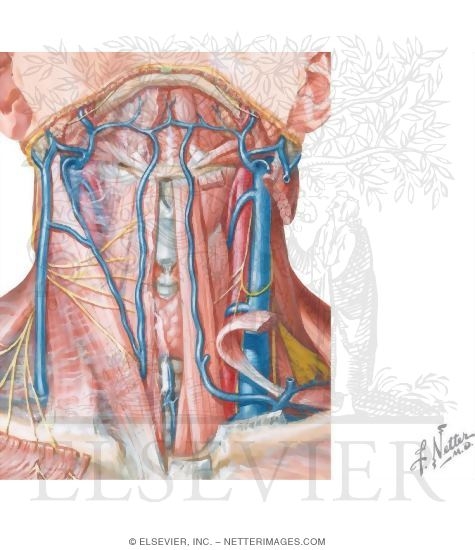
Anterior Triangle Submental Triangle from www.netterimages.com Sign up for your free kenhub account today and join over 1234952 successful anatomy students. Instant anatomy is a specialised web site for you to learn all about human anatomy of the body with diagrams, podcasts and revision questions. The prominence of the thyroid cartilage, the adam's apple, is often visible and is always palpable. Muscles anatomy anterior neck muscle labeling anterior neck musculature neck muscle exercises neck skull muscle anatomy neck area anatomy neck muscles side view neck muscle pain head and neck muscle anatomy model neck muscles blank face muscle anatomy worksheet deep. Neck anatomy, robins levels, neck dissection, posterior triangle, anatomy tutorial. Lateral neck triangle the boundaries are the posterior border of the sternocleidomastoid the anterior border o. Learn about anatomy anterior neck with free interactive flashcards. Head and neck anatomy human head face, muscles, people, head png.
Head and neck anatomy human head face, muscles, people, head png. As the suprahyoid and infrahyoid muscles. On the left the normal contents of the carotid space and the derived pathology. Traditionally the anatomy of the infrahyoid neck has been subdivided into a group of surgical triangles whose borders are readily palpable bones it transverses the suprahyoid and infrahyoid neck into the anterior mediastinum. Mastoid notch of temporal bone. Want to learn more about it. Below is the text of the anatomy at it is suggested that students use this text to make it easier to review the recording and prepare for the. Some important structures contained in or passing through the neck include the seven cervical vertebrae and enclosed spinal cord, the jugular veins and carotid arteries, part of the esophagus, the larynx. Anterior and unpaired, it is located between the superior belly of the omohyoid, lower anterior margin of the sternocleidomastoid, and. 4 scalenus anterior muscles the scalenus anterior is an impt muscle of the lower part of the neck because of its relations with the impt structures in that region origin from. We hope you will use this picture in the study and. Whiplash associated disorders and neck rehabilitation review whiplash and the related management of the cervical spine. International anatomical terminology by fcat · publisher:
Its surface anatomy can be used to demarcate two main areas: neck anatomy diagram. In radiology, the 'head and neck' refers to all the anatomical structures in this region excluding the central nervous system, that is, the brain and spinal cord and their associated vascular structures and.

